

About Lehe
乐于心,和与众,与己乐,与人和; 心宽念纯,百病无生。


乐于心,和与众,与己乐,与人和; 心宽念纯,百病无生。

Today, we will present a case of pancreatic cancer to you. Mrs. Li, 66 years old, is an old patient of ours. Her case was also presented last July (click on the blue text to jump to the reading) . Now, another year has passed, how is she doing now?
01
Case Analysis
If you cannot understand the subsequent professional description,
you can finish reading this text in two minutes.
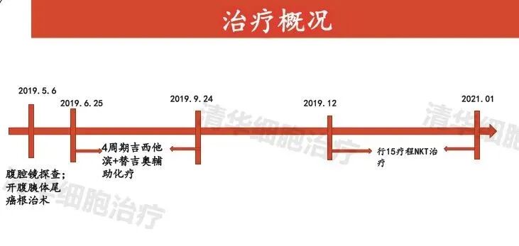
Mrs. Li immediately underwent radical resection of the pancreatic body and tail cancer in May 2019. The pathological results showed moderately-to-poorly differentiated adenosquamous carcinoma, with small foci presenting sarcomatoid structure and focal obvious fibrosis, without definite intravascular thrombus or neural invasion. Immediately afterward, she received four cycles of adjuvant chemotherapy with gemcitabine and temozolomide. After chemotherapy, the results showed a reduction in the accumulation of fluid in the abdominal and pelvic cavity, indicating that the disease had been somewhat controlled.
However, upon reexamination three months later, the MR results revealed suspicious abnormal signals in the residual pancreatic parenchyma, and the abnormal signals in the right lobe of the liver were similar to those previously observed. Meanwhile, the tumor marker CEA gradually increased and exceeded the normal range.
These reexamination results were not ideal, indicating a continued trend of disease progression, and the previous chemotherapy had not effectively suppressed the disease.
Assessment of Mrs. Li's condition
Pancreatic cancer is highly malignant, and the pathological types of patients are complex - "moderately-poorly differentiated", "adenosquamous carcinoma", and there are also "sarcomatoid" structures, posing a high risk of subsequent recurrence and metastasis.

The patient developed an accumulation of fluid in the abdominal and pelvic cavity after surgery. Although it decreased after adjuvant chemotherapy, it still persists and cannot be taken lightly.
The routine follow-up after chemotherapy indicated suspicious signals in the residual pancreas and right lobe of the liver. Considering the elevated tumor marker, the possibility of disease progression in the patient has increased.
NKT cell immunotherapy can delay the time of recurrence and metastasis, and currently represents the optimal time for patients to undergo treatment.
In December 2019, Mrs. Li underwent NKT cell therapy. Due to her high risk of recurrence and metastasis, an intensive regimen of two courses per month was adopted for treatment initially.

Treatment Results
In July 2020, half a year later, the patient completed 8 courses of continuous systemic treatment. Through 3 follow-up visits and imaging examinations, no clear signs of tumor recurrence were found, and the tumor marker CEA decreased to normal values. indicates that the high risk of recurrence and metastasis has been suppressed.
Now that one year of treatment has passed, Mrs. Li, who has completed 15 courses, not only did not show clear signs of tumor recurrence or metastasis on imaging during the re examination in January 2021; The previously elevated CEA has returned to normal levels; The remaining CA125 and CA19-9 are within the normal range.
Science popularization tips
Now, after a year of treatment, her appetite has improved, and her quality of life has significantly increased - her quality of life score has reached 90.5 points, and she even rides a shared bicycle to and from her treatments.
02
Treatment Overview Analysis
>>On May 17, 2019, the pathology report showed: ① (Resected specimens of pancreas, spleen, and left adrenal gland): Based on morphological and immunohistochemical results, the lesion was consistent with moderately-to-poorly differentiated adenosquamous carcinoma, with small foci presenting sarcomatoid structures and focal areas with significant fibrosis. No definite intravascular thrombus or neural invasion was observed, and no cancer was found at the pancreatic resection site. No definite metastasis was observed in the lymph nodes around the pancreas. No definite cancer involvement was found in the spleen and adrenal gland; ② (Resected end of hepatic artery nerve plexus) was examined and found to consist of fibrous adipose connective tissue and nerve fiber bundles, with no cancer observed; ③ (Resected end of celiac trunk mesenteric artery nerve plexus) was examined and found to consist of fibrous adipose connective tissue and nerve fiber bundles, with no cancer observed.
>>From June 25, 2019, to September 24, 2019, four cycles of adjuvant chemotherapy with gemcitabine and temozolomide were administered. On June 18, 2019, a follow-up abdominal CT scan showed: the resection site of the pancreatic body and tail and spleen, as well as the left adrenal gland, had undergone surgery; the fluid accumulation in the abdominal and pelvic cavity had decreased compared to previous scans.
>>On September 25, 2019, CT showed: 1. A few fibrous strands in both lungs; 2. Small nodules in the left lower lobe of the lung. On September 26, 2019, an upper abdominal MR plain scan showed: changes after resection of the pancreatic body and tail and spleen, as well as the left adrenal gland; suspicious abnormal signals in the residual pancreatic parenchyma, with no abnormal high signals on DWI; the abnormal signals in the right lobe of the original liver were similar to those observed previously (June 19, 2019). On September 3, 2019, tumor markers indicated: CEA 8.44ng/ml↑; on September 10, 2019, tumor markers indicated: CEA 6.74ng/ml↑.
03
Imaging data analysis
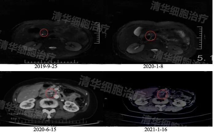
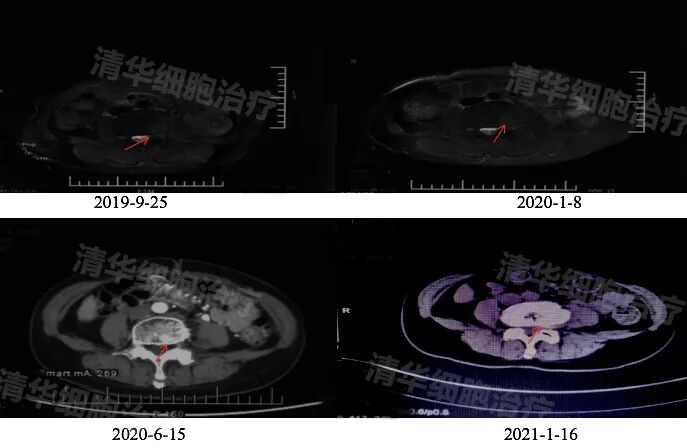
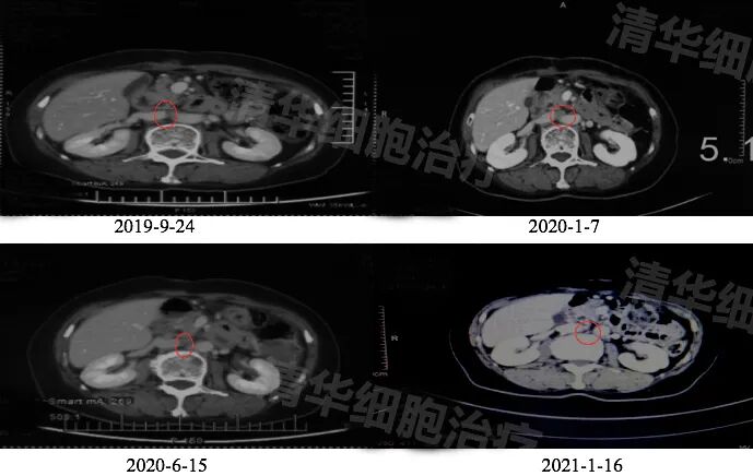
Abdominal MR: Pancreatic body and tail, spleen, left adrenal gland resection postoperative changes, punctate slightly long T2 signal shadow visible within the residual pancreatic parenchyma, size changes not significant from 2019-9 to 2020-1.
Abdominal CT: Compared to 2019-9 and 2020-1, the corresponding high signal area changes were not significant in 2020-6.
Abdominal CT: Compared to 2020-1, there were no significant changes in 2021-1.
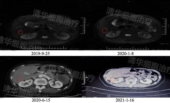
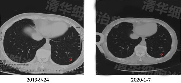
AbdominalMR:Pancreatic body and tail, spleen, left adrenal gland resection postoperative changes, patchy slightly longT2signal shadow visible in the right lobe of the liver, size changes not significant from 2019-9to2020-1.
Abdominal CT: There was no significant change in size between 2020-6 and 2019-9 or 2020-1.
Abdominal CT: There was no significant change in size between 2021-1 and 2020-1 or 2020-6.
04
Tumor marker detection analysis
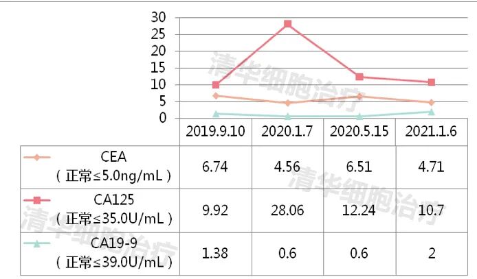
Tumor marker: CEA increased in September 2019, returned to normal in January 2020, and slightly increased in May 2020; CA125 and CA19-9 are within the normal range, with attention to follow-up.
Review of classic NKT cases
Click on the image
to view


Popular science is a kind of charity
<<<Long press to recognize the QR code
Follow Tsinghua Cell Therapy

Address: Room 1101 (Unit 101), 11th Floor, Building 1, No. 7 Courtyard, Kexueyuan Road, Life Science Park, Changping District, Beijing
Phone:010-8265 9866
Email:lehemedneo@hotmail.com